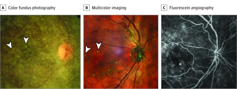
Figure 1. Images by Color Fundus Photography, Multicolor Imaging, and Fluorescein Angiography of Patient 1.
A, A color fundus photograph of the right eye shows asteroid hyalosis; interspersed yellowish spots appear as hard exudate (marked by white arrowheads). B, A multicolor image shows a clear view of the macula, including hard exudates (marked with white arrowheads). C, Fluorescein angiography using the Spectralis system (Heidelberg Engineering) shows multiple leaking microaneurysms.

