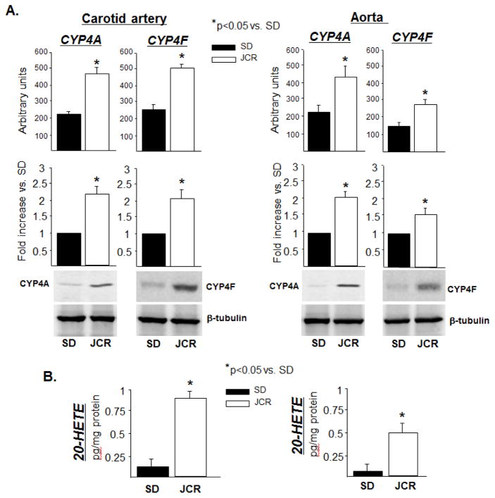
FIGURE 1.
SD and JCR rats were sacrificed and arterial tissue collected. A. Representative Western blots (bottom) and cumulative data (top) showing CYP4A and CYP4F expression in carotid artery and aorta tissue homogenates. B. LC-MS quantitation of 20-HETE production represented in pg/mg protein. n=8, * p<0.05 as indicated.

