
Figure 4. mZFP568 ZF3-10 form base-specific contacts.
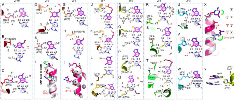
(A–C) ZF10 interacts with a 3-bp triplet (G4-C5-G6). R632 interacts with G4 (A). E629 interacts with C5 (B). R626 interacts with G6 (C).
(D–E) ZF9 interacts with two base pairs (T7-G8). In addition to E601 interaction with T7, S638 at position −5 of ZF10 interacts with A7 of the bottom strand (D). Despite conservation of Ser/Thr at position −5 in each ZF (Figure 1A), the way in which it interacts with DNA differs from finger to finger but most of them interact with the bottom strand.
(F) Superimposition of ZF10 (3-base binder in magenta/red color) and ZF9 (2-base binder in grey color). Note the opposite movement (indicating by green arrows) of base-interacting residues at position −1 and positions −4 and −7 of ZF9.
(G–H) ZF8 interacts with two base pairs (G9-C10). R576 interacts with G9 (G) and E573 interacts with C10 (H).
(I) Superimposition of ZF10 (in magenta/red color) and ZF8 (in grey color) indicates that the small side chain of C570 of ZF8 at position −1 is too far away for direct base contact.
(J–M) ZF7 spans four base pairs (A11-C12-A13-G14). R548 forms a salt bridge with a DNA backbone phosphate group (J). E545, together with T572 of ZF8, interact with the C12:G12 base pair (K). T571 at position −5 of ZF8 interacts with T13 of bottom strand (L). R542 interacts with G14 (M).
(N–Q) ZF6 spans four base pairs (G15-T16-A17-A18). R520 interacts with G15 (N). H517 bridges between T16 and T17 (O and P). S514 interacts with T18 of the bottom strand (Q).
(R–T) ZF5 interaction with three A:T base pairs (A19:T19-A21:T21). L492 interacts with T19 (R). S488 and Q489 interact with T20 (S). Besides C486 and S488 of ZF5, a layer of water molecules separates Y458 and T460 of ZF4 and the A21:T21 base pair (T).
(U–W) ZF3 interacts with three G:C base pairs (G22-G24). R436 interacts with G22 (U). H422 interacts with G23 (V). R430 interacts with G24 (W).
(X) Superimposition of ZF10 (in magenta/red color) and ZF7 (in grey color). Note the different conformations of two arginine residues at positions −1 and −7.
(Y) H517 bridges between two base pairs 15 and 16.