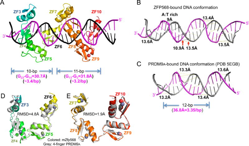
Figure 5. DNA and protein conformational changes.

(A) After removing ZF1-2, ZF3-10 could be divided into two 4-finger fragments with each contacting 10 or 11 base pairs.
(B) mZFP568-bound DNA has a narrower minor groove spanning the A:T rich sequence.
(C) For comparison, PRDM9A-bound DNA has a consistent minor groove width (PDB 5EGB).
(D–E) Superimposition of 4-finger array of PRDM9A with that of ZF3-6 (D) and ZF7-10 (E). See also Figure S5 and Table S3.
