Fig. 3.

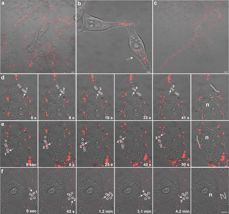
Representative confocal images at low magnification of a Vero cells with Au@DynPro b HEK293T cells with Au@ShortPro and c MDCK cells with Au@TransRb. Representative images showed similar NP dispersion throughout the culture and cell-to-cell transfer of Au@DynPro through short (b) or long (c) projections. d, e Representative time-lapse images of the linear progression of Au@DBP inside the cell and the resulting trajectories (circles). f Comparison with the non-linear movement obtained with control NPs Au@IntCt. Scale bar: 5 µm
