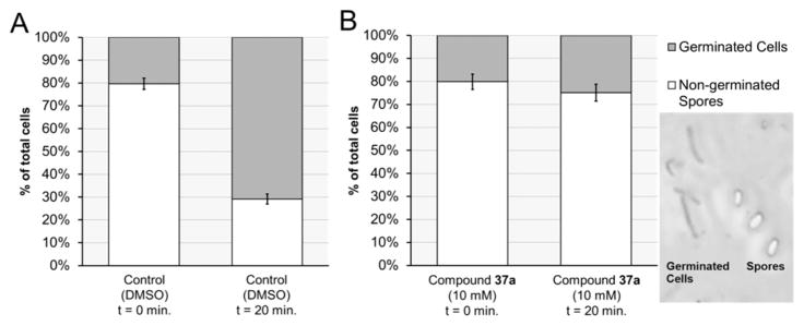
Figure 7.
Cell count using phase-contrast microscopy to determine percentage of spore germination over 20 min. (A) Under the conditions of the control experiment (BHIS, 2000 μM TCA, and DMSO), the number of germinated spores rose from 20% to 71% over 20 min. (B) In contrast, far fewer spores germinated under the same conditions in the presence of 10 μM of 37a (from 20% to 25%). Inset: Phase-contrast microscope image of germinated cells (phase dark) and spores (phase bright).

