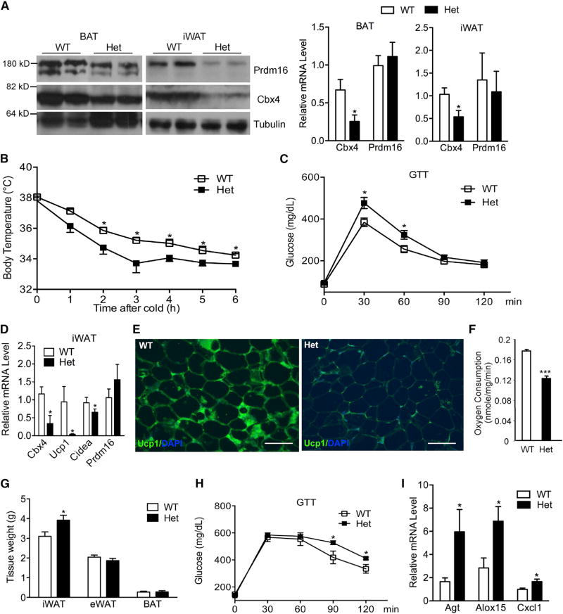
Figure 6. Cbx4 Haploinsufficiency Causes a Reduction of Prdm16 Protein and Impaired iWAT Browning, Leading to iWAT Expansion and Deteriorated Glucose and Cold Tolerance.
(A) Analysis of Prdm16 and Cbx4 protein and mRNA (n = 4 mice/group) from 8-week-old male Cbx4-heterozygous mice and control littermates. Data represent mean ± SEM. *p < 0.05.
(B) Body temperature of 10-month-old male Cbx4-heterozygous mice (n = 8 mice) and control littermates (n = 7 mice) at 4°C. Data represent mean ± SEM. *p < 0.05.
(C) Glucose tolerance test in a second cohort of 10-month-old male Cbx4-heterozygous mice (n = 6 mice) and control littermates (n = 8 mice). Data represent mean ± SEM. *p < 0.05.
(D) Gene expression in iWAT of 10-month-old male Cbx4-heterozygous mice and control littermates (n = 4 mice/group). Data represent mean ± SEM. *p < 0.05.
(E) Ucp1 immunofluorescence staining of iWAT of 10-month-old male mice. Shown are representative images of three mice per genotype. Scale bar, 200 µm.
(F) Oxygen consumption of iWAT isolated from 8-month-old male Cbx4-heterozygous mice and control littermates (n = 5 mice/group). Data represent mean ± SEM. ***p < 0.001.
(G) Fat mass of male Cbx4-heterozygous mice (n = 7 mice) and control littermates (n = 5 mice) on a high-fat diet for 25 weeks. Data represent mean ± SEM. *p < 0.05.
(H) Glucose tolerance test of mice in (G) after 24-week high-fat diet. Data represent mean ± SEM. *p < 0.05. (I) Inflammation gene expression in iWAT of mice in (G). *p < 0.05.

