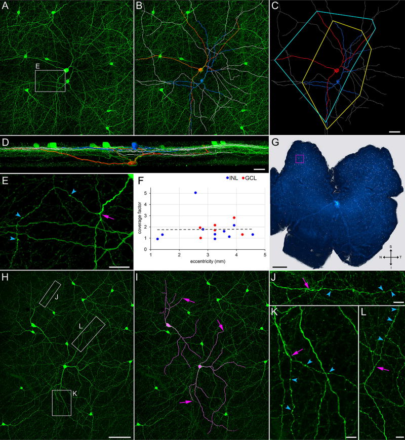
FIGURE 2.
Coverage factors. Flat-mounted retina (G) immunostained for TH (green). (A–D) Field marked by magenta square in G. (A) Z-projection (thickness = 71.5 µm) of optical sections through the INL, IPL, GCL, and nerve fiber layer. (B) Superimposition, on A, of tracings of (in blue) a TH cell soma in the INL and its tapering neurites, (in red) a TH cell soma in the GCL and its tapering neurites, and (in gray) the varicose neurites of these cells. (C) Tracings with no background, together with polygons that connect the tips of the tapering neurites. (D) 90 ° rotation of B. (E) Higher magnification display of field outlined by rectangle (labeled E) in A, showing termination (arrow) of a tapering neurite and emergence of a varicose neurite as a side branch. (F) Coverage factors measured for cells with soma in the INL (blue dots) or GCL (red dots), at eccentricities 1 to 5 mm from optic nerve head in a total of six different retinae. Optic nerve head in G is light area near center of retina. (H) TH cells at an eccentricity of 3.25 mm in the inferior–temporal quadrant of a different retina than G. (I) Tapering neurites of two of these cells are highlighted in magenta. (J–L) Varicose neurites extending beyond the tip of these tapering neurites, within the areas outlined by the correspondingly lettered boxes in H. Arrows in I (and arrows at corresponding positions in J–L) point at end of tapering portions, and beginning of varicose portions, of neurites. Arrowheads point at a few varicosities in each neurite (E, J–L). Scale bar = 50 µm in C (applies to A–C); 30 µm, 25 µm, and 1 mm in D, E, and G, respectively; 100 µm in H (applies to H,I); 10 µm in J–L

