FIGURE 7.
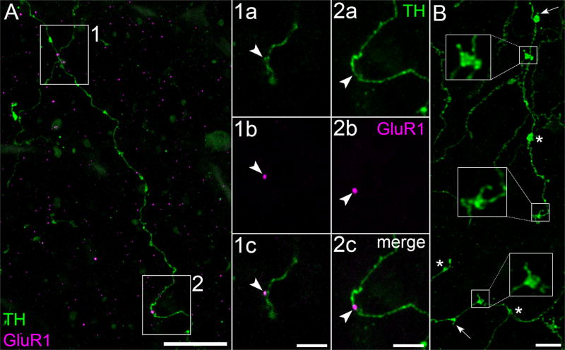
GluR1 and spine-bearing varicosities in OPL. (A) Portions of TH cell varicose neurite (green) in the OPL of flat-mounted retina. Z-projection (thickness = 9.2 µm). Two neurites criss-cross in box 1. Panels 1a and 2a show the boxed areas in A at higher magnification. Panels 1b and 2b show one of the optical sections from these panels with only the magenta color channel (GluR1) turned on. Panels 1c and 2c merge the TH and GluR1 signals, showing GluR1 on the shaft of a varicose neurite (panel 2c) and on a small, apparently stubby spine (panel 1c). Arrowheads point to identical positions in the panels of each column. (B) Varicose neurites of TH cell (green) in the OPL. The large boxes show higher magnifications of the varicosities outlined in the small boxes, and thin spines attached to each outlined varicosity. Arrows point to other spine-bearing varicosities. Asterisks are next to spine-free varicosities. Scale bar = 20 µm in A; 5 µm in 1c,2c,B

