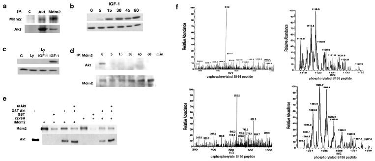
Figure 2.
Phosphorylation of Mdm2. (a) Western blot analysis of insulin receptor (control, 2 μg), Akt, and Mdm2 (2 μg) immunoprecipitated from serum-starved MCF-7 cells probed with anti-Mdm2 (IF-2) or anti-Akt. (b) Serum-starved MCF-7 cells were stimulated with 50 ng/ml IGF-1 and a Western blot was probed with anti-phospho (activated)-Akt (Upper). The blot was stripped and reprobed with anti-Akt (Lower). (c) Serum-starved MCF-7 cells were treated with medium or LY294002 for 30 min and then IGF-1 for 30 min. Western blots were probed with anti-phospho Akt (Upper) or anti-Akt (Lower). (d) MCF-7 cells were stimulated with insulin before immunoprecipitation of Mdm2. A Western blot was probed with anti-Akt and anti-Mdm2. (e) Recombinant Mdm2 or 2xSA was incubated with GST or GST-Akt-agarose for 30 min in vitro, centrifuged, and washed three times. Vehicle or raAkt was incubated with the agarose complexes for 30 min at 37°C and these were centrifuged and washed three times. A Western blot probed with anti-Mdm2 or anti-Akt quantitated the amount of Mdm2 or 2xSA that had associated with GST-Akt agarose. (f) Mdm2 immunoprecipitated from MCF-7 cells incubated with vehicle or IGF-1 was analyzed by mass spectrometry. Predicted parent ions for unphosphorylated serine 166 and serine 186 were recovered from the control incubates (Left), whereas parent ions predicted for phosphorylated serine 166 and serine 186 were recovered from cells treated with IGF-1 (Right).

