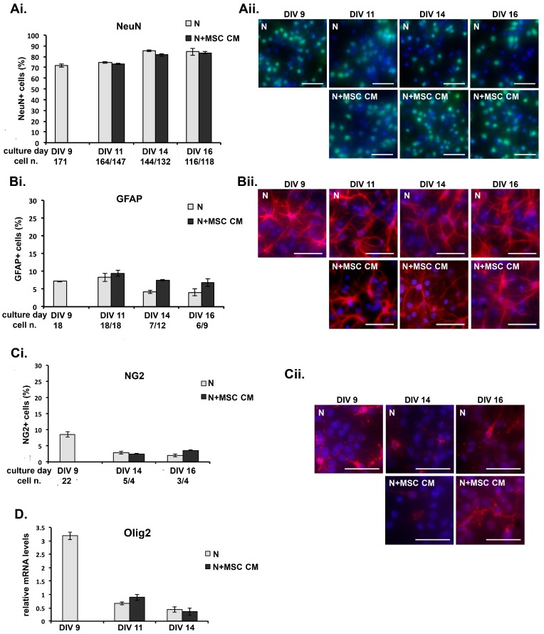
Figure 3.
MSC secreted factors do not induce differential survival of neurons or expansion of non-neuronal cells in long-term cultures. (A–C) DIV 9 cortical neurons were grown in MSC CM for different lengths of time, fixed and immunostained using antibodies for cell markers, (A) neuronal nuclei (NeuN) for neurons, (B) glial fibrillary acidic protein (GFAP) for mature astrocytes and new neurons, and (C) neural/glial antigen 2 (NG2) for oligodendrocyte precursor cells (OPC), microglia/macrophages and pericytes, and nuclei counterstained with the fluorescent DNA stain 4′,6-diamidino-2-phenylindole (DAPI). The proportions of each cell type (percentage of specifically-immunostained cells compared to total numbers of DAPI-stained cells) in the untreated and MSC CM-treated cultures at each time point are shown (Ai, Bi, Ci). The total numbers of DAPI-stained cells/field in cultures at each time point and condition are shown under the x-axis. Representative fields from each culture are shown in photographs (Aii, Bii, Cii). Scale bars 100 μM. (D) DIV 9 cortical neurons were grown in MSC CM for different lengths of time, and levels of mRNA for Olig2 measured by quantitative PCR. Results shown are means ± SEM of triplicate samples from one representative of three independent experiments. p > 0.05 for pairwise comparisons by Student’s t-test.

