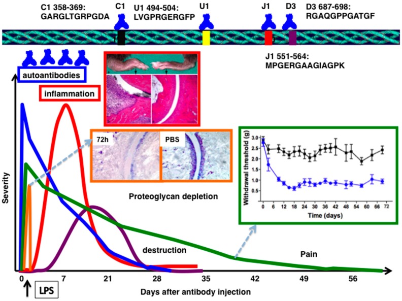
Figure 1.
Schematic diagram of acute form of collagen antibody induced arthritis. Autoantibodies binding to well defined epitopes are transferred at day 0, followed by injection of lipopolysaccharide from E. coli 05:B55 as the secondary stimulus at day 3. Significant level of proteoglycan depletion was observed 72 h after antibody injection. Inflammation (red and swollenness) and, cartilage and bone erosions between arthritis and control mouse are shown. HE stained joint morphology of arthritis and control mice. Magnification, 10×. Pain (withdrawal threshold levels) started even before inflammation began and prolonged even after resolution of inflammation. Dotted arrows indicate the inserted figures.

