Figure 3.
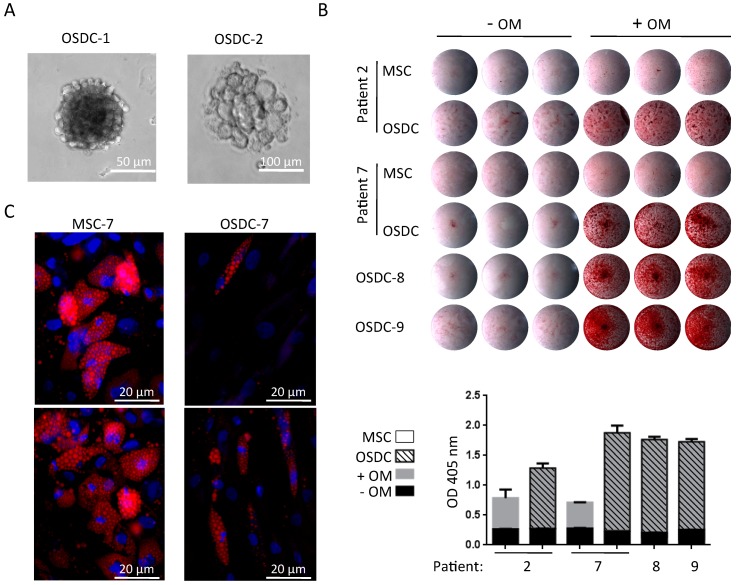
Osteosarcoma derived cells (OSDC) in anchorage-independent, osteogenic or adipogenic culture conditions. (A) Optical microscopy observation of sphere clones in anchorage-independent culture conditions; (B) Calcium deposition observed with alizarin red staining following 21 days of cell culture with (+OM) or without (-OM) osteogenic induction media. Images of stained-culture wells are shown for OSDC or MSC plated in triplicate for each culture condition. Bound alizarin red was solubilized and optical density (OD) was measured for each well. The histogram represents mean values with standard deviation of each triplicate; (C) Representative images of MSC and OSDC from patient 7 following 14 days of cell culture with adipogenic induction medium are shown. Adipocytes containing small Nile Red-positive lipid droplets are easily observed (red vesicles) whereas undifferentiated cells were not labeled with Nile Red. Nuclei were counterstained (blue color) with 4′,6-diamidino-2-phenylindole (DAPI).

