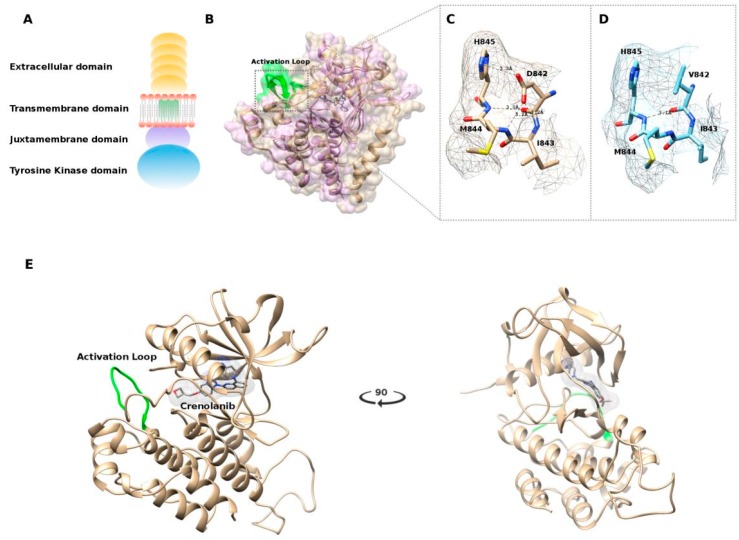
Figure 5.
(A) Schematic representation of the platelet-derived growth factor receptor alpha (PDGFRA) receptor; (B) Structure alignment performed using the jCE algorithm, between the crystallized structure of the c-Kit–Imatinib complex (PDB: 1T46) in purple and the structure of PDGFRA (PDB: 5K5X) in gold. Highlighted in green is the Activation loop; (C) Polar interactions of the 842 residue with wild-type Aspartic Acid; (D) Polar interactions of the 842 residue carrying the mutated Valine amino acid; (E) Representation of the best docked pose of crenolanib in the PDGFRA ATP binding site.

