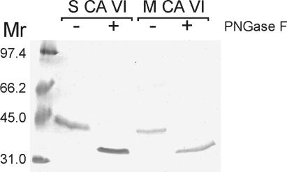
Figure 3.
PNGase F treatment of human salivary and milk CA VI followed by SDS/PAGE and Colloidal Coomassie blue staining. Without PNGase F treatment (−), the 42-kDa polypeptides for both salivary (S) and milk (M) CA VI are seen, corresponding to the glycosylated form of CA VI, but after digestion (+), the 36-kDa polypeptides for both samples are seen, indicating that the two glycopolypeptides have polypeptide cores of similar sizes.

