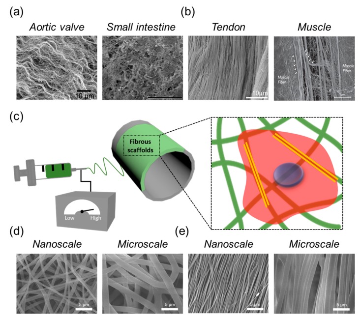
Figure 1.
Scanning electron microscope (SEM) images of a natural extracellular matrix (ECM) in distinct types of tissues with (a) isotropic direction and (b) anisotropic direction [30,31,32,33]; (c) Schematic illustration of the electrospinning process; (d) Representative SEM images of fibrous scaffolds with a controllable fibrous scale with (d) randomly and (e) aligned fibrous deposition via electrospinning.

