Figure 4.
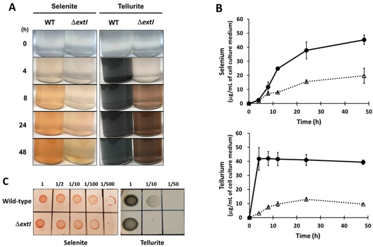
The effects of extI deficiency on selenite and tellurite reduction. (A) The colorimetric change observed during selenite and tellurite reduction. Wild-type (WT) and extI-deficient mutant (∆extI) cells were anaerobically cultured in FWA medium containing 500 µM selenite (left panels) or 500 µM tellurite (right panels) as only the electron acceptors; (B) wild-type (circle and solid line) and ∆extI (triangle and dotted line) cells were anaerobically cultured in FWA medium containing 500 µM selenite (upper panel) or 500 µM tellurite (lower panel) as the sole electron acceptor. The amounts of selenium and tellurium precipitated were measured by HG-AFS. Experiments were conducted independently two times and error bars represent standard deviation; (C) selenite and tellurite susceptibility. The wild-type and ∆extI cells were anaerobically cultured in NBAFYE medium. After adjusting for bacterial OD, cells were diluted as indicated with saline and spotted onto the NBAFYE agar plate with 500 µM selenite (upper panel) and 500 µM tellurite (lower panel), and then cultured for 3 days.

