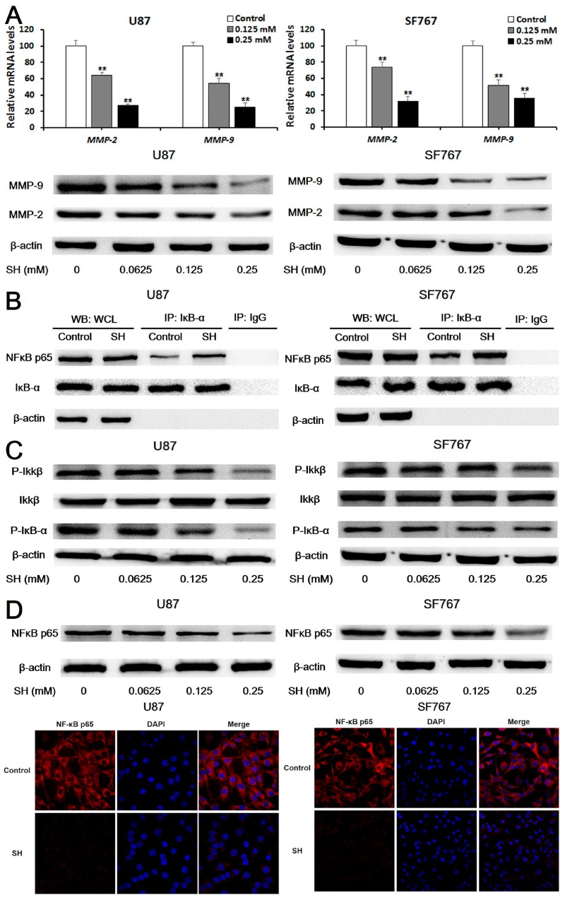
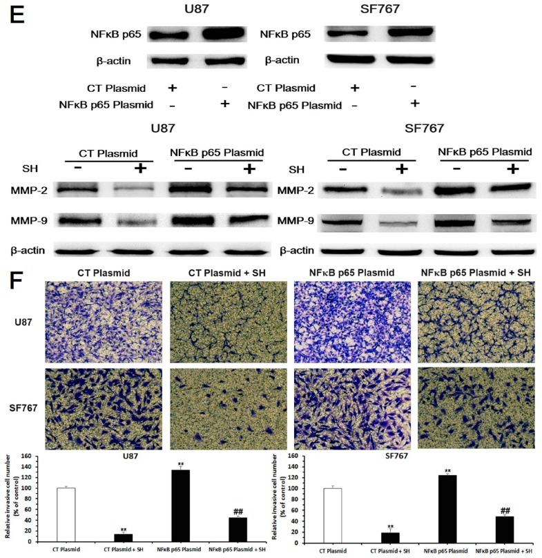
Figure 3.
SH reduced matrix metalloproteinase (MMP)-2/-9 expression by suppressing nuclear factor kappa B (NFκB) activation. (A) After treatment with the indicated concentrations of SH for 24 h, levels of the MMP-2/-9 mRNAs in U87 and SF767 cells were assessed by quantitative real-time PCR, and levels of the MMP-2/-9 proteins in the two cell lines were examined using Western blot analysis; (B) Co-immunoprecipitation (Co-IP) analysis of the binding of NFκB p65 and inhibitor of NFκB (IκB)-α. Cells were incubated with SH (0.25 mM) for 6 h. An IκB-α antibody was used in the IP experiment, an NFκB p65 antibody was used in the subsequent Western blot analysis, and an IgG antibody was applied as the negative control. WCL: whole cell lysates; (C) The SH treatment decreased levels of phosphorylated inhibitor kappa B kinase (IKK) β and IκB-α. U87 and SF767 cells were incubated with the indicated concentrations of SH for 24 h; (D) SH inhibited NFκB p65 expression in U87 and SF767 cells. Prior to the Western blot analysis, cells were treated with the indicated concentrations of SH for 24 h. For the immunofluorescence assays, cells were treated with SH (0.25 mM) for 24 h, fixed, stained with primary antibodies against NFκB p65, and stained with fluorescein isothiocyanate (FITC)-labeled secondary antibodies (red fluorescence). Nuclei were stained with diamidino-phenyl-indole (DAPI) (blue fluorescence). The protein was observed using a laser scanning confocal microscope, the images of U87 cells were captured at 1000× magnification and the images of SF767 cells were captured at 400× magnification; (E) Efficiency of NFκB p65 overexpression and the effects of an NFκB p65 plasmid on SH-induced down-regulation of MMP-2/-9 expression. Cells were transfected with the no-target control plasmid (CT plasmid) or the NFκB p65 plasmid for 24 h and then treated with or without SH (0.25 mM) for another 24 h; (F) Effects of the NFκB p65 plasmid on the SH-mediated reduced invasion of the two cell lines in matrigel-coated Transwell invasion assays (100× magnification). Cells were transfected with the CT plasmid or the NFκB p65 plasmid for 24 h and then treated with or without SH (0.25 mM) for an additional 24 h. Each image is representative of n = 3 experiments. All blots shown above are representative of n = 3 experiments with similar results. β-Actin served as the loading control. All data are presented as means ± SEM, n = 3. ** p < 0.01 compared with the control; ## p < 0.01 compared with the NFκB p65 plasmid-transfected group.


