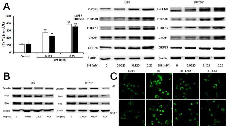
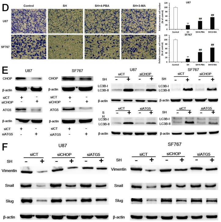
Figure 4.
SH impairs the invasiveness of human glioblastoma cells by reversing epithelial-mesenchymal transition (EMT) through endoplasmic reticulum (ER) stress-mediated autophagy. (A) Effects of SH on [Ca2+]i and levels of ER stress-associated proteins in U87 and SF767 cells. Cells were treated with the indicated concentrations of SH for 24 h; (B) Effects of SH on the protein levels of mesenchymal markers in the two cell lines. Cells were treated with the indicated concentrations of SH for 24 h; (C) Twenty-four hours after SH (0.25 mM) treatment in the presence or absence of 4-phenylbutyric acid (4-PBA) or 3-methyladenine (3-MA), the two cell lines were stained with monodansylcadaverine (MDC) and photographed with a laser scanning confocal microscope (600× magnification); (D) Treatments with 4-PBA or 3-MA compensated for the SH (0.25 mM)-mediated inhibition of the invasiveness of the two cell lines at 24 h (100× magnification); (E) Efficiency of CCAAT/enhancer binding protein (C/EBP) homologous protein (CHOP) or autophagy-related 5 (ATG5) knockdown and the effects of CHOP or ATG5 silencing on the SH-induced increase in the microtubule-associated protein light chain 3B (LC3B)-II levels. Cells were transfected with a no-target control siRNA (siCT), CHOP siRNA (siCHOP) or ATG5 siRNA (siATG5) for 24 h and then treated with or without SH (0.25 mM) for an additional 24 h; (F) Effects of CHOP or ATG5 silencing on the SH-mediated decreased protein levels of mesenchymal markers. Cells were transfected with siCT, siCHOP or siATG5 for 24 h and then treated with or without SH (0.25 mM) for an additional 24 h. Each blot and image shown is representative of n = 3 experiments. Data represents means ± SEM, n = 3. ## p < 0.01 compared with the SH treatment alone; ** p < 0.01 compared with the control.


