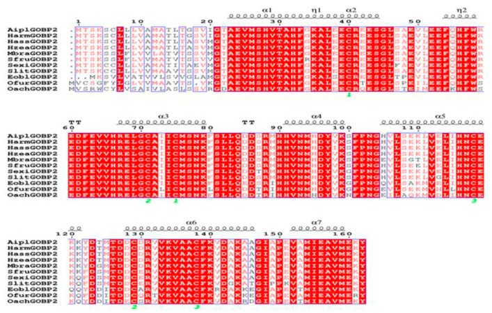
Figure 1.
Multiple sequence alignment of 11 GOBP2 homologous proteins sequences. Green numbers represent six conserved cysteines in the GOBP2s. Strictly identical residues are highlighted in white letters with red background. Residues with similar physicochemical properties are shown in red letters. Alignment positions are framed in blue if the corresponding residues are identical or similar. The secondary structure elements for GOBP2s are shown on the top of the sequences; α-helices are displayed as wave line.

