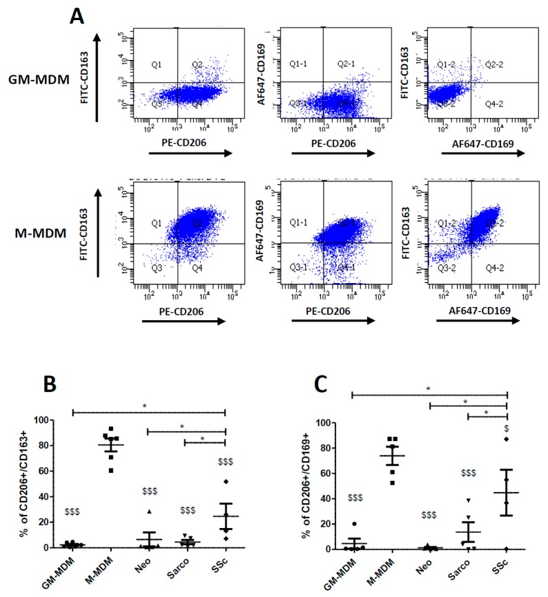
Figure 3.
Comparison of the percentage of cells co-expressing CD206, CD163 and CD169 between GM-CSF and M-CSF-derived MDMs and AM from patients suffering of lung neoplasia, sarcoidosis or SSc-ILD. Primary human monocytes from healthy donors were differentiated into MDMs in vitro in the presence of GM-CSF (GM-MDM) or M-CSF (M-MDM) for 6 days. Bronchoalveolar lavage fluids of patients were washed and cells were plated until the following day. Cells were then harvested, stained and the expression of cell surfaces molecules was analyzed by flow cytometry. Graphs representing the percentage of GM-CSF or M-CSF-derived MDMs co-expressing CD206/CD163, CD206/CD169 or CD163/CD169 are representative of 5 independent experiments (A). Data expressed as the percentage of CD206+/CD163+ cells +SEM (B) or of CD206+/CD169+ cells +SEM (C) are the means of 5 independent experiments except for SSc-ILD with 4 samples. ANOVA followed by Newman–Keuls’ multiple comparison Test: * p < 0.05; $ p < 0.05 and $$$ p < 0.001 when compared to M-MDM.

