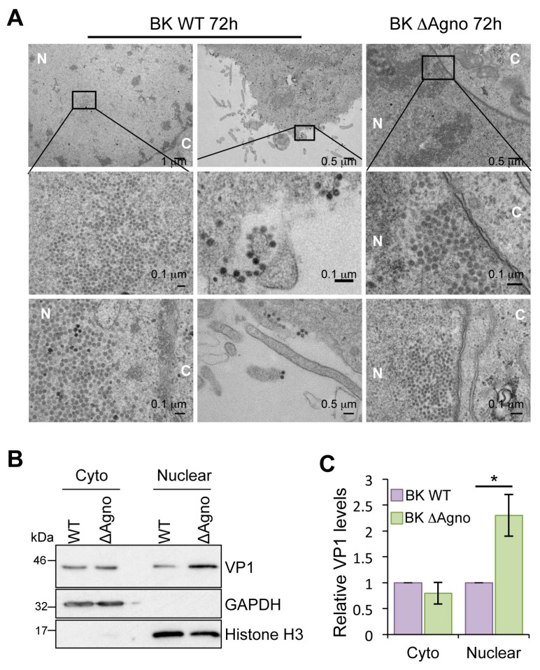
Figure 4.
Agnoprotein facilitates nuclear release of BK virions. (A) Electron microscopy analysis of BK WT and ΔAgno infected RPTE cells (n = 40 cells). Boxed areas in the upper panel are shown at higher magnification in the middle panels. Viral particles of about 40 nm in diameter were found in the nuclei of BK WT and ΔAgno transfected cells. Nuclei (N) and cytoplasm (C) are labeled. Scale bars are shown in the panels; (B) Cell fractionation of RPTE cells transfected with BK WT or ΔAgno genomes. Fractions were probed with for VP1 expression. Antibodies detecting GAPDH and Histone H3 served as markers for the cytoplasm and nuclear fractions; (C) Quantification of the Western blot data was performed using ImageJ software (1.8.0_101, NIH, USA) on the VP1 positive bands and is represented relative to BK WT VP1. The graph corresponds to an average from three independent experimental repeats. Significance was analyzed by Student’s t-test and is indicated by an asterix * p <0.05.

