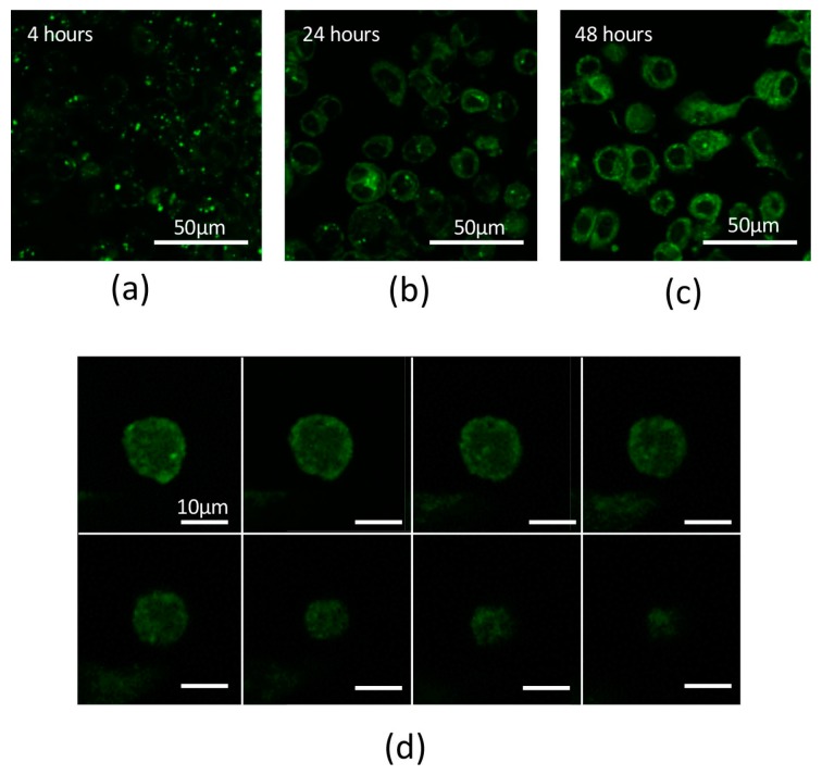
Figure 3.
(a) Confocal laser scanning microscopy (LSM) image showing strong Alexa 488 fluorescence intensity on the surface of SK-BR3 cells. PCSN probes were bound to the surface of SK-BR3 cells within 4 h. (b) LSM image showing strong fluorescence signals inside the SKBR3 cells. PCSN probes were internalized into the cytoplasm during a 24-h incubation in medium containing targeted PCSN probes (1200 ppm). (c) PCSN probes remained inside the SK-BR3 cells after 48 h. (d) For three-dimensional analysis of the SK-BR3 cells, images were collected at 0.41-µm intervals with a 488-nm laser to create a Z stack. The images show internalized PCSN probes inside an SK-BR3 cell.

