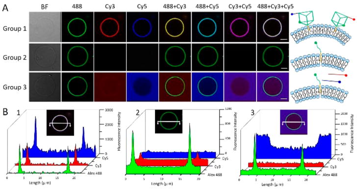
Figure 2.
Anchoring DNA nanoprisms on cell-mimicking giant vesicles. (A) Confocal laser scanning microscopy imaging of colocalization of 500 nM nanoprisms on MVs. Panel 1: TP-Cy3 and TP-Cy5 anchored on lipid bilayer through cholesterol-insertion. Panel 2: Single strand of Chol-DNA-Alexa Fluor 488. Panel 3: Single strand of Chol-DNA-Alexa Fluor 488 and free arm strands of Cy3-DNA and Cy5-DNA. All samples were incubated with 400 μL solution of MVs at 37 °C for 30 min. Scale Bar: 5 μm. (B) Cross section of fluorescence intensities (white solid line) in Groups 1, 2 and 3, respectively.

