Figure 3.
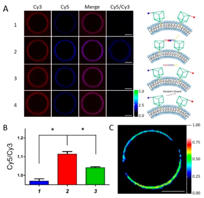
Assembly/disassembly of 3D DNA nanostructures on cell-mimicking membrane of MVs. (A) Confocal laser scanning microscopy imaging of manipulation of DNA nanoprisms on MVs in 1640 medium containing 5 mM Mg2+ at 4 °C. (1) 500 nM TP-Cy3 and TP-Cy5. (2) Linker strand added to form dimeric nanoprism assembly of two separate TPs. (3) Displacement strand added to disassemble the dimeric nanoprism. (4) Random linker strand added to group (1) showed little effect on assembly. (B) Normalized fluorescence intensity measurements for panels 1 to 3 from panel A. Each column represents the statistical sample population of 15 MVs. P values were calculated by Newman–Keuls Multiple Comparison Test, *P < 0.05. (C) Acceptor bleaching experiment to study FRET efficiency on an individual MV. Scale bar: 5 μm.

