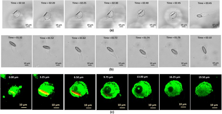
Fig 2. Internalization of fabricated tag structures into the melanoma skin cancer cells.
The time sequence images of two internalization events for two tag sizes: (a) 9 μm × 15 μm and (b) 9 μm × 21 μm. (c) The Z-sectional images of the fluorescent labeled cells and tag with the depth of focus varies by 1 μm, the tag size is 9 μm × 18 μm.

