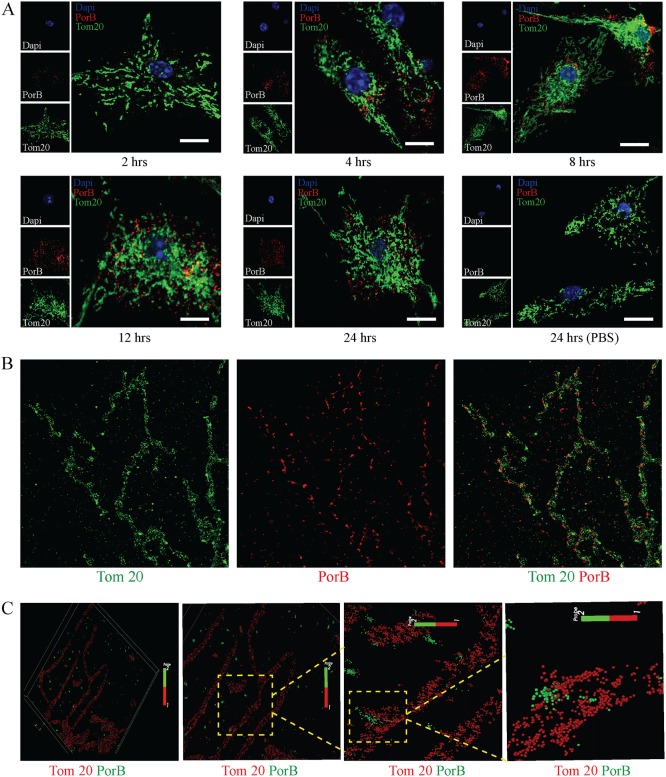
Fig 5. OMVs enable mitochondria targeting of PorB.
(A) Bone marrow-derived macrophages (BMDMs) were treated with purified N. gonorrhoeae OMVs and analyzed by confocal laser scanning microscopy for PorB (red) and Tom20 (green) localization at indicated times. Cells were stained with DAPI (blue) to visualize nuclei. Scale bar = 10 μm. Representative images of more than 200 cells from three biological samples. (B) BMDMs treated with purified OMVs for 12 hours were analyzed by RapidSTORM reconstructed dual colour super-resolution imaging for PorB (red) and Tom20 (green) localization. (C) OMV treated BMDMs (12 hours) were probed with anti-PorB (green) and Tom20 (red) antibodies and analyzed by 3D single-molecule localization super-resolution microscopy.

