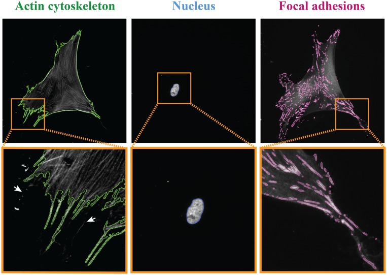
Fig 4. Detection of a single cell, nucleus and focal adhesions (FAs).
Representative grey-scale images of the actin cytoskeleton, nucleus, and FAs of HVSCs on a substrate homogeneously coated with fibronectin. The detected outlines are shown in green, blue, and magenta, respectively, and the orange rectangles marked areas show zoom-in images of the cell, nucleus and FAs. The white arrows indicate some small actin-rich membrane protrusions that were not detected.

