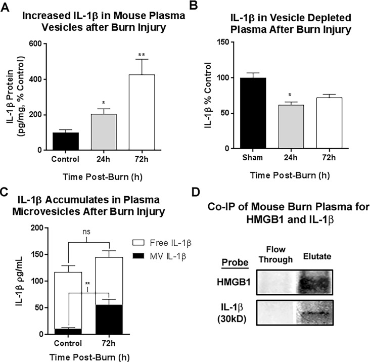
Fig 2. IL-1β is concentrated in mouse plasma microvesicles (MVs), but not vesicle depleted plasma up to 72 hours after burn injury.
Mice underwent a 20% TBSA thermal injury and were sacrificed at either 24 or 72 hours post-burn. Plasma microvesicles (MVs) were isolated from by sequential centrifugation. IL-1β levels were assessed by ELISA in the MVs and MV-depleted plasma. (A) IL-1β was increased in plasma MVs up to 426% of control levels. At 24 hours post-burn IL-1β was 2-fold control levels; 203.1±32.1 vs. 100±10.7% control, mean±SEM, Burn vs Control, group *p<0.05, N = 3 control, 4 burn. At 72 hours post-burn IL-1β levels in MVs were 400% control values; 426.1±88.09 vs. 100 ± 10.7% control, Burn vs Control, mean±SEM, **p<0.01, N = 5 per group. Data from two separate experiments were combined and presented as percent control. (B) In MV-depleted plasma, IL-1β levels were not increased. At 24 hours, MV-free IL-1β was significantly reduced by 21%; 100±8.43% vs 79.12±4.02%, mean±SEM, Control vs Burn, p<0.05, N = 4–5. At 72 hours post burn a nonsignificant reduction in IL-1β was observed; 100±10.10% vs 80.43±12.36%, mean±SEM, Control vs Burn, p = 0.24, N = 5–7. (C) Analysis of plasma fraction, MV versus MV free plasma, showed that plasma IL-1β increases at 72 hours were due to increases in MV fraction. (D) Co-immunoprecipitation (Co-IP) of mouse plasma for HMGB1 was performed. Column flow through and eluate were assessed by western blot for HMGB1 and IL-1β. Both HMGB1 and IL-1β were found in the eluate indicating HMGB1 and IL-1β complex formation in mouse plasma. Both the 31kD pro-form of IL-1β and HMGB1 were detected in the eluate, but not the flow through demonstrating the presence of HMGB1/pro-IL-1β complexes in human plasma.

