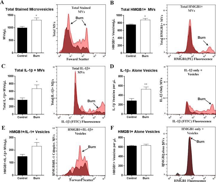
Fig 3. HMGB1 and IL-1β Co-localize in plasma microvesicles (MVs) following burn injury.
Mice underwent a 20% TBSA thermal injury and were sacrificed at 72 hours post-burn. Plasma microvesicles (MVs) were isolated from by sequential centrifugation. Vesicles were permeabilized using Fix/Perm buffer and labeled with anti-HMGB1 and anti-IL-1β antibodies. MVs between 0.1–1.0μm were identified using MegaMix size gating beads. Positive staining was differentiated from background by comparing to single color controls for each antibody. (A) The total number of stained MVs was increased by 34% at 72 hours after burn injury; **p<0.01, N = 8 control, 6 burn. (B) The total number of HMGB1+ MVs was increased by 17%; 753.5 ± 19.6 vs 879.1 ± 41.7; Control vs Burn N = 7 Control, 6 Burn (C) The total number of IL-1+MVs was increased by 78%; 377.7 ± 54.9 vs 672.5 ± 99.1 Control vs Burn N = 8 Control, 6 Burn. (D) MVs positive for IL-1β alone were increased after burn injury by 61%; 281.9 ± 51.22 vs 536.6 ± 45.76, Control vs Burn, *p<0.05, N = 7 control, 5 burn. (E) MVs positive for both HMGB1 and IL-1β were increased by 64%; 145.4 ± 15.30 vs 237.8 ± 32.74, Control vs Burn, *p<0.05, N = 7 control, 6 burn. (F) MVs positive for HMGB1 alone were not changed after burn injury, 612.7 ± 13.27 vs 641.3 ± 22.73, Control vs Burn, p = 0.27, N = 8 control, N = 6 burn. Representative relative frequency histograms depicting different MV populations for each panel are included: red-burn, white-control. **p<0.01, p<0.05, t-test vs control.

