Fig. 4.
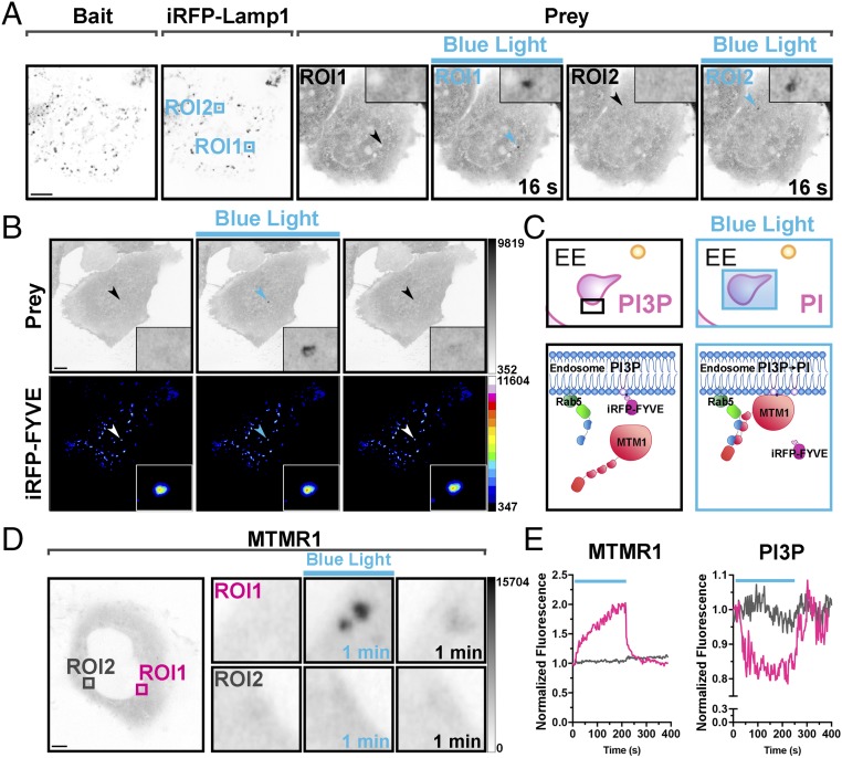
Light-dependent recruitment of a prey protein at single-organelle resolution. (A) Localized protein recruitment to a single lysosome. HeLa cell expressing Lys-nMag (bait), pMagFast2(3x)-tgRFPt (prey), and iRFP-Lamp1 (lysosomal marker). iRFP fluorescence was used to select the area of blue-light illumination (arrows). Blue boxes indicate two areas subjected to sequential 200-ms blue-light pulses at 0.5 Hz for 2 and 1 min, for ROI1 and ROI2, respectively. The prey is recruited selectively to the illuminated lysosome as shown (Inset). (Magnification: Insets, 5×.) (Scale bar: 10 μm.) (B) Localized protein recruitment to a single endosome. HeLa cell expressing nMag-Rab5 (not shown), pMagFast2(3x)-mCherry (prey), and iRFP-FYVE [PI3P binding endosome marker]. iRFP fluorescence was used to select the endosome to be illuminated (arrows). (Insets) Prey recruitment at that endosome. (Magnification: Insets, 6×.) (Scale bar: 10 μm.) (C–E) Selective reduction of PI3P levels on a single endosome (magenta box in the left micrograph of D), without affecting PI3P on the other endosomes (e.g., gray box), by the specific recruitment of the PI3P phosphatase MTMR1 to that endosome. Schematic cartoon depicting the experiment is shown in C, and a quantification of the recruitment to endosomes of MTMR1 (mCherry fluorescence) and of the levels of PI3P (iRFP fluorescence) on the same endosomes is shown in E. (Magnification: D, Right Insets, 7×.) (Scale bar: 5 μm.)

