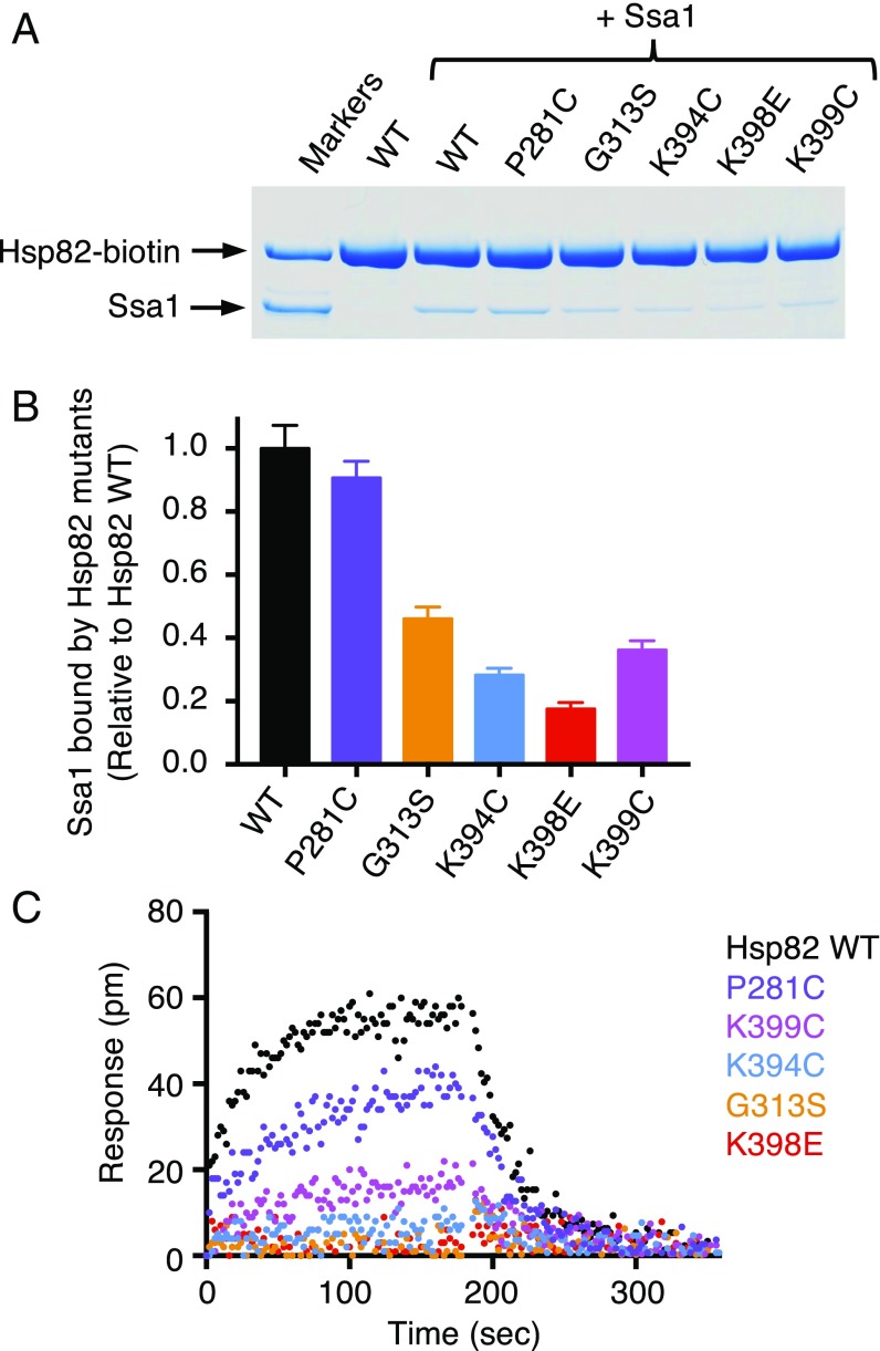
Fig. 5.
Hsp82 mutant proteins exhibit defective interaction with Ssa1 in vitro. (A) The interaction between biotin-labeled wild-type or mutant Hsp82 (1 μM) and Ssa1 (4 μM) was measured in a pull-down assay as described in Materials and Methods. The proteins were analyzed by SDS/PAGE and visualized by Coomassie staining; the gel shown is a representative of at least three independent experiments. (B) Quantification of Ssa1 bound to wild-type or mutant Hsp82-biotin from A. The ratio of Ssa1 bound to each of the biotin-labeled Hsp82 mutants relative to wild-type Hsp82 is presented as mean ± SEM. (C) The kinetics of association and dissociation for the interaction between 60 μM Ssa1 and biotinylated wild-type (black) or mutant (colored) Hsp82 was monitored by BLI as described in Materials and Methods. Representative curves are shown for three or more independent experiments.

