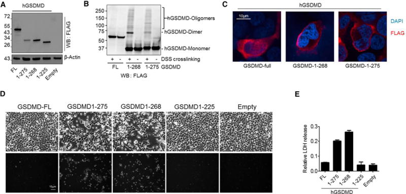
Figure 7. ELANE Cleaves GSDMD to Generate a Smaller but still Biologically Active GSDMD-eNT Fragment.

(A) Expression of FLAG-tagged FL, ELANE-cleaved (hGSDMD-eNT or 1–268), and caspase-cleaved (hGSDMD-cNT or 1–275) human GSDMD in HEK293T cells. The cells were lysed 24 hr post-transfection.
(B) GSDMD protein oligomerization. The cells expressing the indicated hGSDMD protein were lysed with lysis buffer with or without disuccinimidyl suberate crosslinking reagent (1 mg/mL) and incubated at room temperature for 30 min. The reaction was quenched by 0.1 M Tris, pH 7.4.
(C) Subcellular localization of recombinant hGSDMD in 293T cells. Shown are representative images of three independent experiments.
(D) Cell morphology of transfected cells was observed by bright field microscopy 24 hr post-transfection (see also Movies S1, S2, S3, and S4). PI staining was conducted to assess cell death. Shown are representative images of three independent experiments.
(E) Cytotoxicity was measured using a lactate dehydrogenase (LDH) cytotoxicity assay. All of the values represent means ± SDs of three independent experiments.
