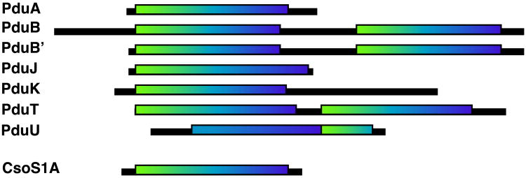
Figure 7.
Cartoon representations of the seven S. enterica Pdu shell proteins containing BMC domains recognizable by sequence homology. The boxes outline the sequence segments containing predicted BMC domains. The typical BMC domains are shaded from white at the N-terminus to black at the C-terminus. PduU can be aligned to parts of two consecutive BMC domains, or can alternatively be described as a circular permutation of a single BMC domain.

