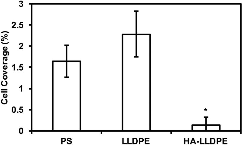
Figure 6.
(a) Representative fluorescence microscopy images of adhered platelets and leukocytes stained with Calcein-AM on PS, LLDPE, and HA-LLDPE surfaces.
(b) Percent coverage analysis of adhered platelets and leukocytes after staining with Calcein-AM on PS, LLDPE, and HA-LLDPE surfaces. Percent coverage of platelets were determined by the evaluation of 15 representative images taken per group over 3 studies (n = 45) using ImageJ. Each image covered an area of 25 µm2. Results indicated significantly less cellular adhesion on HA-LLDPE as compared to other surfaces (p ≤ 0.05).


