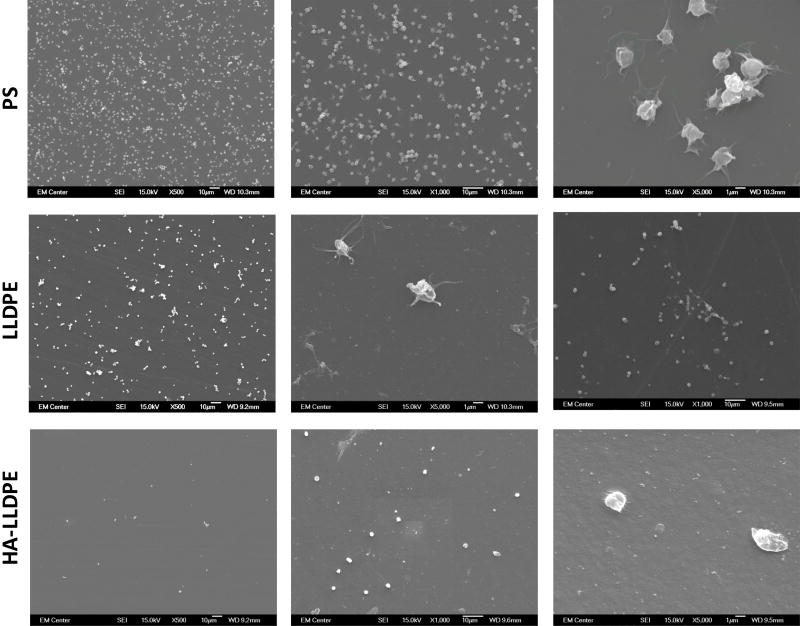
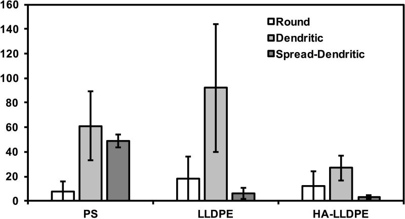
Figure 7.
(a) Representative SEM images of adhered platelets and leukocytes on PS, LLDPE, and HA-LLDPE surfaces. Results indicate HA-LLPE surfaces promoted no activation of platelets as noted by the rounded morphology. By comparison PS and LLDPE both resulted in fully activated platelets as expressed by the spread-dendritic morphology of platelets on these surfaces.
(b) Platelet count on different surfaces. Qualitative SEM analysis was preformed using the methods described elsewhere on 15 representative images per group taken over 3 studies at 1000C (n = 45). Each image covered an area of 144 µm2. For HA-LLDPE, the number of un-activated platelets was significantly lower than dendritic and dendritic-spread platelets (p ≤ 0.05).


