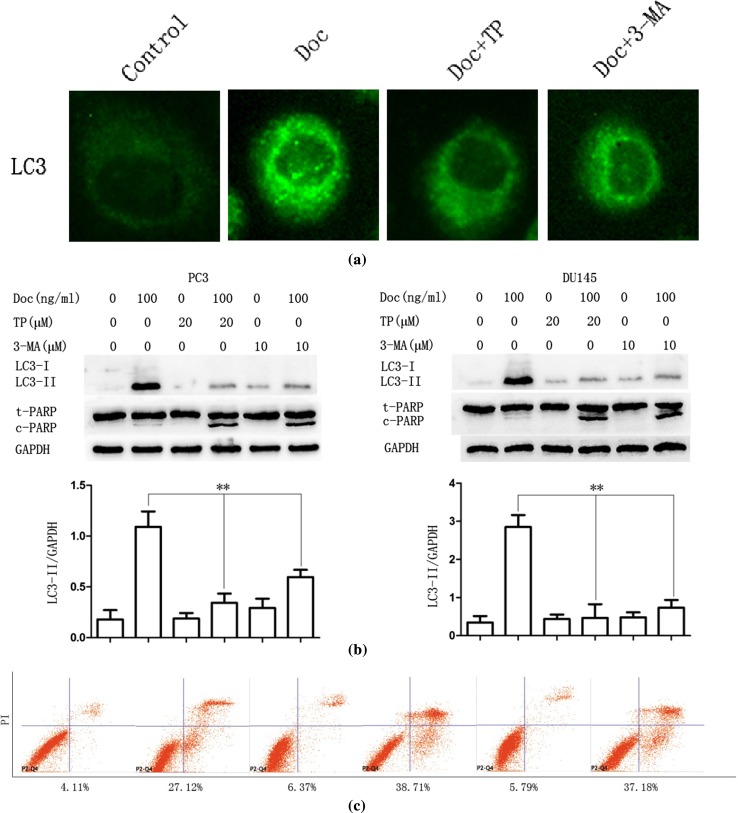
Fig. 3.
TP inhibits docetaxel-induced autophagy and promotes apoptosis in PC3 and DU145 cells. a PC3 cells were cultured in Doc (100 ng/ml) for 12 h with TP (20 μM, 30 min) or 3-MA (10 μM, 30 min) pretreatment, LC3 punctate formation was assayed by confocal microscopic analysis. Images are representative of 10 random fields. b PC3 and DU145 cells were cultured in Doc (100 ng/ml) for 24 h with TP (20 μM, 30 min) or 3-MA (10 μM, 30 min) pretreatment, cell extracts were analyzed for protein expression using western blot analysis. c PC3 cells were cultured in Doc (100 ng/ml) for 24 h with TP (20 μM, 30 min) or 3-MA (10 μM, 30 min) pretreatment, and cell apoptosis was measured by flow cytometry (FCM). The percentages of early and terminal stage apoptotic cells and necrotic cells were calculated. *P < 0.05, **P < 0.01

