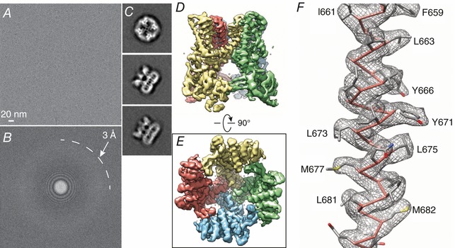
Figure 3. The first high resolution cryo‐EM structure of an ion channel (Liao et al. 2013).

A, cryo‐electron micrograph of TRPV1. Micrographs typically have low contrast. B, Fourier transform of micrograph shown in A showing high resolution information approaching 3 Å. C, representative 2D class averages of particles. D and E, 3D density map of TRPV1 with each subunit colour‐coded, viewed from side (D) and bottom (E). F, cryo‐EM density map of the TRPV1 S6 domain superimposed on the atomic model. The clearly defined geometry of α‐helices and side‐chain densities allowed de novo model building. Reproduced with permission from Nature Publishing.
