
Figure 2. Shank3 isoforms improve the surface expression of recombinant HCN2 channel in Xenopus oocyte.
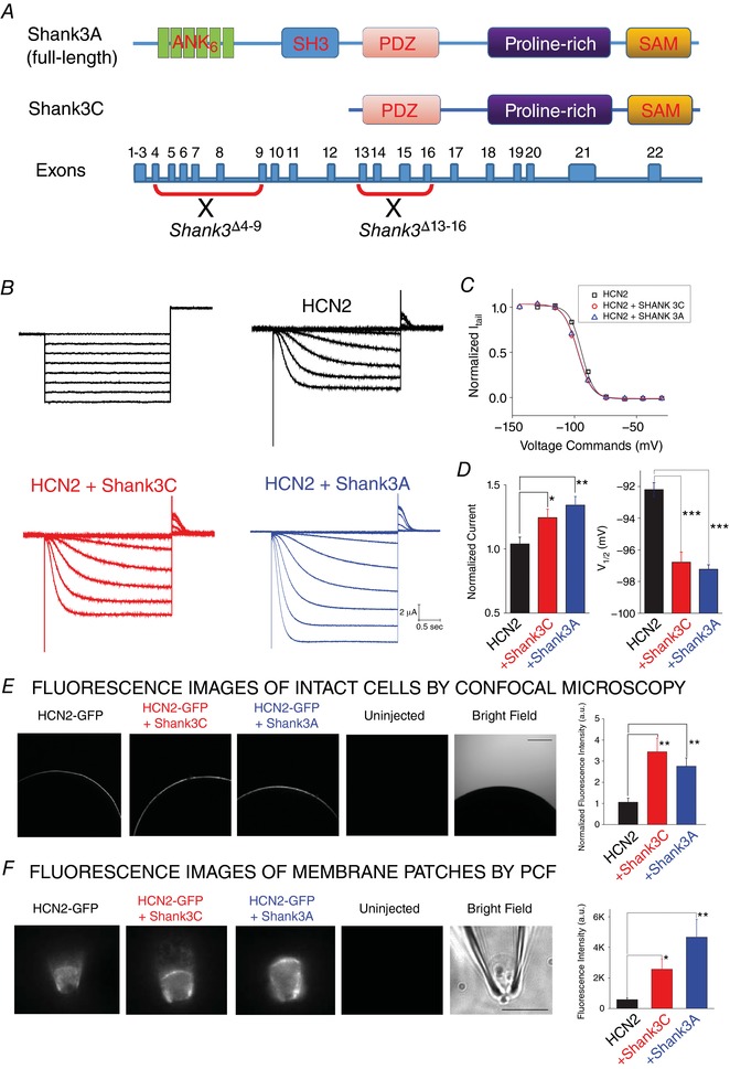
A, schematic representation of the genomic structure of Shank3 gene and two splicing isoforms, Shank3A (full‐length) and Shank3C (missing ankyrin and SH3 domains in the N‐terminus). B, representative HCN2 current (top; no Shank3) recording from whole oocytes by TEVC. A series of hyperpolarizing voltage steps (bottom) were used to activate the channel. C, HCN2 current recorded from oocytes co‐injected with Shank3A (top) or Shank3C (bottom). D, summary of Shank3 effects on the function of HCN2 channel. Values of V 1/2 (mV): HCN2, −92.2 ± 1.1; HCN2 + Shank3C, −96.0 ± 1.5; HCN2 + Shank3A, −97.2 ± 0.6. n = 6 for each group. E, confocal images of whole oocyte expressing (from left to right) HCN2 alone, HCN2 + Shank3A, HCN2 + Shank3C, and uninjected cell. F, fluorescence images of membrane fractions pulled from oocyte surface and held within the glass patch‐clamp pipette in the inside‐out configuration. Statistics are shown on the right. Unpaired t test: * P < 0.05; ** P < 0.01; *** P < 0.001. [Color figure can be viewed at http://wileyonlinelibrary.com]