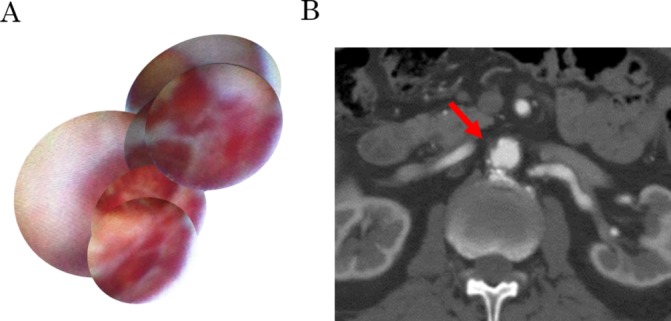
Figure 2.

A large cavity 2 years later at the site of the subintimal haemorrhage. (A) Angioscopic image. A larger cavity with a complicated structure and frames covered with red thrombi were seen. (B) Axial CT image of the corresponding area. Slightly progressed hazy intimal thickening was found without any sign of aortic dissection or obvious penetrating atherosclerotic ulcer.
