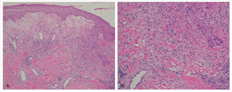
Figure 2.
(A) Papillary dermal oedema with an inflammatory infiltrate of the superficial to mid dermis, involving the perivascular and interstitial regions (x100 magnification). (B) Superficial and mid dermal perivascular and interstitial lymphohistiocytic inflammatory infiltrate with neutrophils and leucocytoclasis (x200 magnification).

