Figure 2.

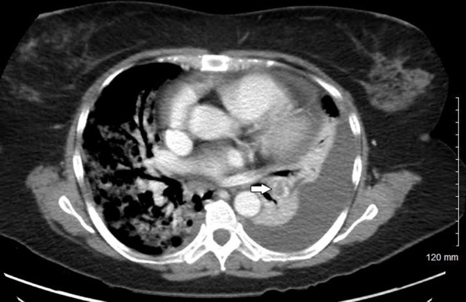
Perfusion defect due to a pulmonary embolus in the lobar artery to the left lower lobe (white arrow). Left pleural effusion with compression atelectasis can also be seen.

Perfusion defect due to a pulmonary embolus in the lobar artery to the left lower lobe (white arrow). Left pleural effusion with compression atelectasis can also be seen.