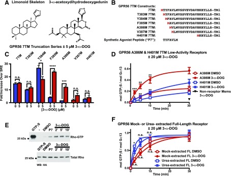
Fig. 2.
3-α-Acetoxydihydrodeoxygedunin is a small-molecule activator of GPR56/ADGRG1. (A) Chemical structure of 3-α-acetoxydihydrodeoxygedunin (3-α-DOG) side-by-side with a generalized skeleton for the limonoid class. (B) Comparison of the tethered-agonist regions of engineered GPR56 7TM receptors. (C) GPR56 7TM receptors with intact (7TM) or truncated tethered-agonist residues were treated with vehicle (blue bars) or with 5 μM 3-α-DOG (red bars) in an SRE-luciferase gene reporter assay. Data are normalized to Renilla luciferase control and expressed as fold over the signal obtained from cells transfected with SRE-luciferase only. Error bars are the mean ± S.D. of three biologic replicates with two technical replicates per experiment. (D) GPR56-A386M 7TM (truncated tethered-peptide-agonist) and GPR56-H401M 7TM (no tethered-peptide-agonist) receptor membranes, or control, nonreceptor membranes were reconstituted with purified, recombinant Gα13 and Gβ1γ2. Membranes were preincubated with 3-α-DOG or DMSO for 5 minutes. [35S]GTPγS binding to G13 was then measured over time. Graphs were fitted to monoexponential functions using GraphPad Prism. Error bars are the mean ± S.D. of three experimental replicates with two technical replicates per experiment. 3-α-DOG-mediated activation of A386M 7TM and H401M was significant compared with control, DMSO-treated receptor (*P < 0.05; **P < 0.01; ***P < 0.001; ****P < 0.0001). (E) RhoA activation by acute treatment of GPR56-A386M 7TM-expressing HEK293T cells with 3-α-DOG or P7 synthetic peptide agonist. The Western blot is representative of three independent experiments. (F) Untreated and urea-treated GPR56 full-length membranes were reconstituted with Gα13 and Gβ1γ2 and preincubated with 0 or 20 μM 3-α-DOG prior to measurement of G13 [35S]GTPγS binding kinetics. Error bars are the mean ± S.D. of three experimental replicates with two to three technical replicates per experiment (*P < 0.05; n.s., not significant compared with cells treated with DMSO).

