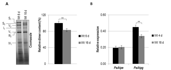
Figure 1. FIGURE 1: Age-dependent dimer decrease and analysis of expression of PaAtpe and PaAtpg in P. anserina.

(A) Representative BN-PAGE analysis and quantification of mitochondrial protein extracts (V2, F1Fo-ATP-synthase dimers) from three independent 6-days and 16-days old wild-type strains. F1Fo-ATP-synthase dimers were normalized to Coomassie stained gel. Relative dimer content of 6-days old wild-type strains was set to 100%. Mitochondrial protein complexes were stained with Coomassie Blue and complexes and supercomplexes S1, S0, V2, I1, V1, III1 and IV1 are indicated.
(B) Transcript analysis of PaAtpe and PaAtpg in three young (4 d) and old (16 d) wild type cultures, respectively. RNA was isolated and gene expression was determined by qRT-PCR analyses. The relative expression was normalized to the expression of PaPorin coding for a mitochondrial outer membrane protein. Error bars represent the standard deviation and the P-values were determined by Student´s t test.
