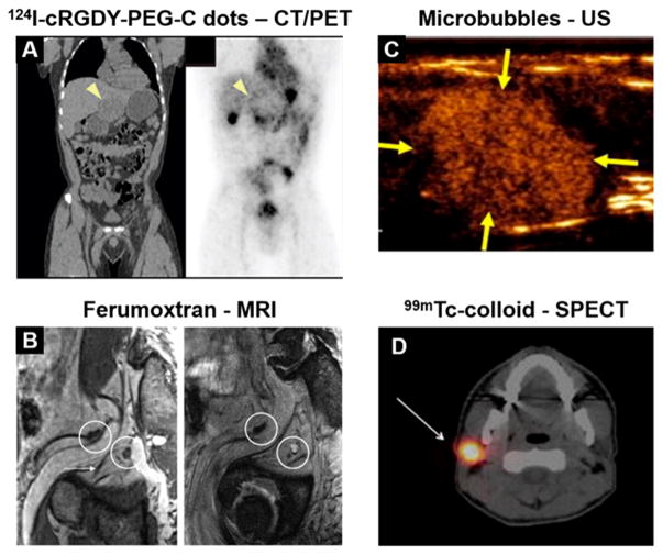
Figure 1.
Clinical examples of NP imaging agents. (A) Administration of 124I-cRGDY-PEG-C dots shows accumulation of the tracer near the periphery of a hepatic lesion. Reprinted with permission from ref 33. Copyright 2014 AAAS. (B) Ferumoxtran-enhanced MRI enables visualization of a metastatic lymph node, which appears hyperintense in a T2*-weighted image postcontrast (right) due to its lack of NP uptake. Reproduced with permission from ref 13. Copyright 2009 RSNA Publications. (C) Targeted microbubbles provide contrast of a breast lesion using ultrasound. Reprinted with permission from ref 50. Copyright 2017 American Society of Clinical Oncology. All rights reserved. (D) 99mTc-colloid preferentially accumulates in a sentinel lymph node. Reproduced with permission from ref 42. Copyright 2017 Springer.

