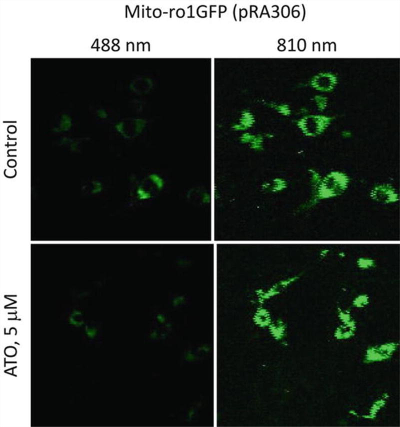
Fig. 2.
SK-Mel-28 melanoma cells stably expressing a mito-ro1GFP plasmid pRA306 were examined by a laser confocal microscope at 528 nm for green fluorescence. Top panels: no-treatment control cells, bottom panels: 5 µM arsenic trioxide (ATO) treated for 24 h. Left panels: excited at 488 nm; right panels: excited at 810 nm

