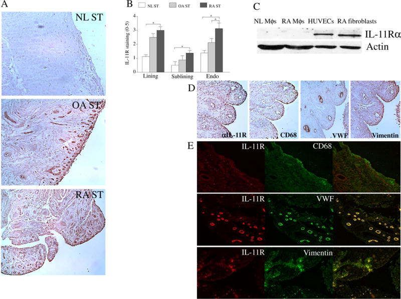
Fig. 2. IL-11Rα expression is markedly higher in ST lining fibroblasts and sublining fibroblasts and endothelial cells of RA compared to NL ST.

A. STs from NL, OA and RA were stained with anti-IL-11Rα Ab (original magnification x 200) and (B) staining was scored on a scale of 0–5 in the lining, sublining and endothelial cells, n=8−9. C. The IL-11Rα expression was quantified in NL and RA PB in vitro differentiated macrophages (MΦs), RA ST fibroblasts and HUVECs by Western blot analysis using anti-IL-11Rα Ab, n=5. D. RA ST serial sections were stained with Abs to anti-IL-11Rα, CD68, VWF or Vimentin to establish which cell types express IL-11Rα, n=5. E. To confirm serial section studies, individual (red or green) as well as the overlapping (yellow) immunofluorescence staining was shown of RA STs that were stained with Abs to anti-IL-11R (green) or cell markers per slide including CD68 (red), VWF (red) or Vimentin (red) (original magnification x 400), n=5. Values are the mean ± SE. * represents p <0.05.
