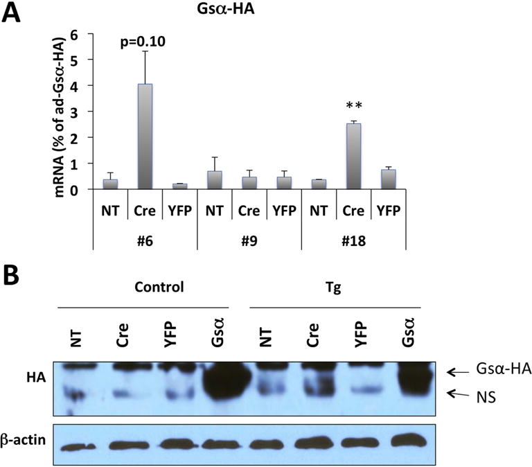
Figure 2. qRT-PCR and Western blot analysis of Gsα-R201H expression in BMSCs.

(A) qRT-PCR analysis of Gsα-R201H mRNA level in BMSCs upon adenovirus transduction. BMSCs were isolated from 4-6-week-old F1 or F2 generation of 3 founder lines. Primers were designed to detect the transgene-derived mutant Gsα transcript by taking advantage of the sequences of the HA tag. mRNA levels were normalized against the samples transduced with adenovirus-Gsα-HA. Data represent mean ± SEM of two-to-three independent experiments; **, p<0.01. (B) Western blot analysis of protein lysates from BMSCs upon adenovirus transduction. BMSCs were isolated from 4-6-week-old F1 or F2 generation cGsαR201H line #6 or control littermates. The Gsα-R201H protein was detected using an antibody against the HA tag; note the immunoreactivity above the non-specific (NS) band in the sixth lane from left. Cells transduced with ad-Gsα-HA were used as positive control. Ad-YFP was used as negative control. NT, non-transduced. β-actin immunoreactivity was used to ensure comparable gel loading.
