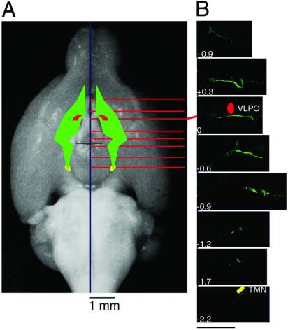
Figure 4.
Schematic representation of the location of DP reconstructed from serial coronal sections. (A) The DP-concentrated region in the ventral surface of the basal forebrain is schematically represented by the green color with emphasis on its topological relation with the ventrolateral preoptic area (VLPO, red) and tuberomammillary nucleus (TMN, yellow). (B) Typical coronal sections are shown in rostral and caudal directions from bregma. The values represent the distance from bregma (mm). (Bar, 800 μm.)

