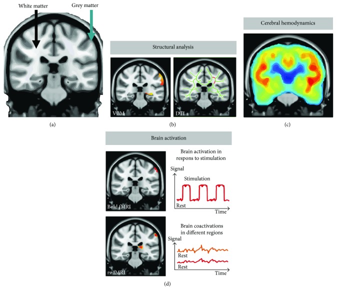
Figure 2.
A brief overview of MRI modalities to elucidate cerebral pathophysiology in relation to POCD and POD. (a) Anatomical MRI scan showing white matter and grey matter of the brain. (b) (Left side) Voxel-based morphometry (VBM) analyses the anatomical images to determine possible changes in grey matter volume and morphometry. (b) (Right side) Diffusion tension imaging (DTI) can characterize white matter integrity and tracts. These modalities can potentially reveal even minor neuroplasticity. (c) Measurement of cerebral hemodynamics with intravenous (IV) contrast permits determination of, for example, cerebral blood flow (CBF), cerebral blood volume (CBV), and cerebral oxidative metabolic rate (CMRO2), while CBF also can be measured without IV contrast by arterial spin labeling (ASL). (d) Functional MRI can indirectly determine neuronal activation by measuring concomitant changes in blood flow. Blood-oxygenation-level dependent (BOLD) fMRI signal is useful in investigating brain activation to an explicit task. Resting state fMRI (rsfMRI) can reveal coactivation of distinct regions across the brain in patients that are not performing an explicit task.

